
What Is the Retroperitoneum? Anatomy, Organs and Functions Explained
Authored by:
Dr Gaurav Aggarwal (Uro-oncologist and Robotic Surgeon)
When patients come to see me with kidney problems, prostate issues, or urological conditions, I often need to explain where things are located inside their body. That’s when I start talking about the retroperitoneum. Most people have never heard of it, and honestly, that’s understandable. It’s not an organ you can point to or something you think about unless something goes wrong.
But here’s the thing—understanding what is the retroperitoneum and why it matters has helped many of my patients grasp why their condition needs the treatment it does. So let me walk you through it.
What Is the Retroperitoneum, Actually?
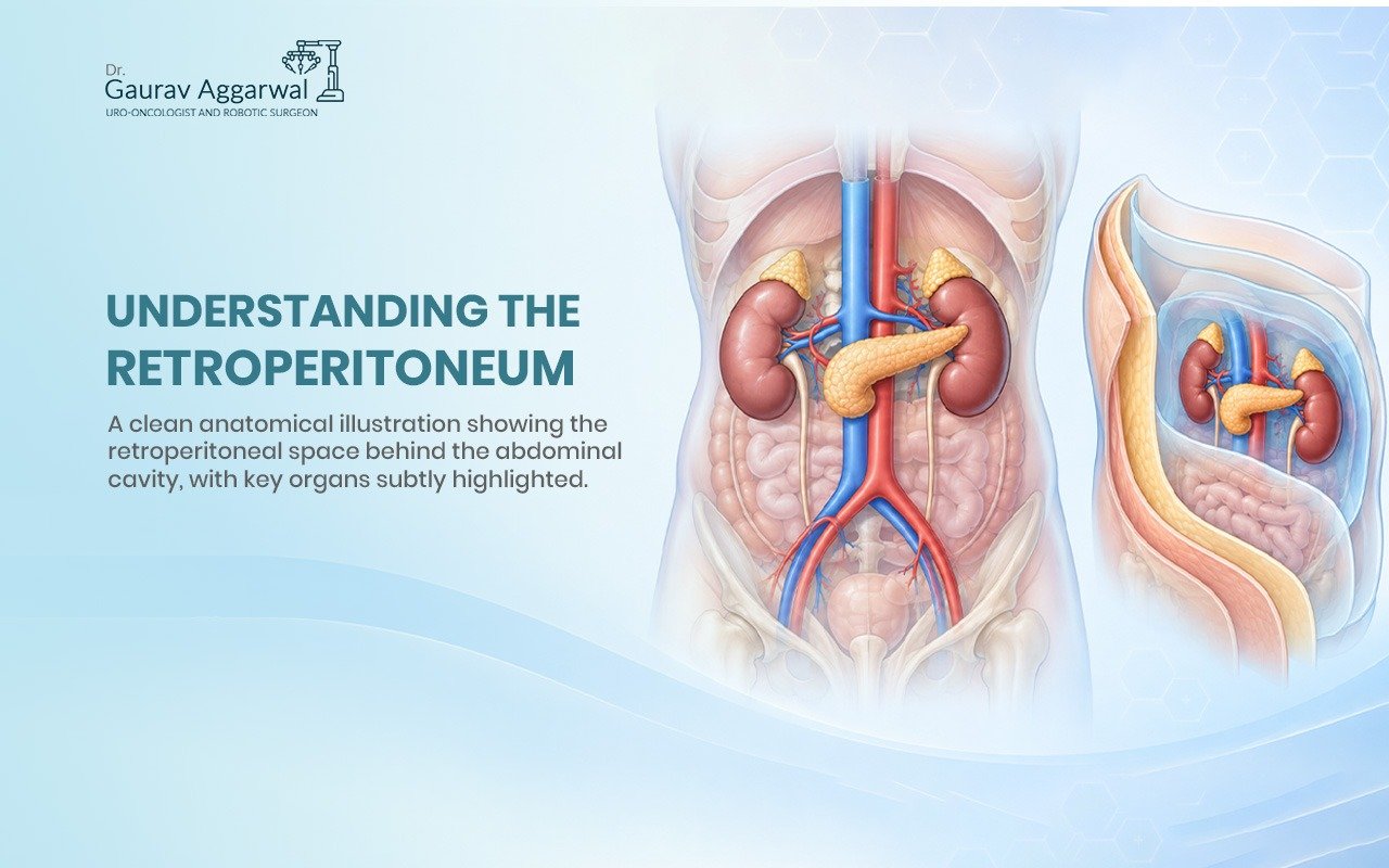
The retroperitoneum is a space. Not an organ, not a tissue—a space. Specifically, it’s the area behind your abdominal cavity that sits between the peritoneum (a membrane lining your abdomen) and the back wall of your abdomen.
Think of it this way: inside your belly, you have your intestines, stomach, liver, and other organs. These organs have a protective lining around them called the peritoneum. Everything inside that lining is called the peritoneal space. But behind that lining—behind the peritoneum—there’s another space. That’s the retroperitoneum.
When I perform robotic surgery on the kidneys or prostate, I’m often working in this retroperitoneal space. This is where the important structures hide, which is why understanding the anatomy matters.
What Organs Are in the Retroperitoneum?
This is where it gets clinically interesting. Several vital organs live in this space, and they’re crucial to your body’s function.
The kidneys sit in the retroperitoneum on either side of your spine, one on the left and one on the right. These filter your blood and produce urine. As a urologist, the kidneys are central to what I treat.
The ureters run from your kidneys down to your bladder, carrying urine. They travel through the retroperitoneum the entire way.
The adrenal glands sit on top of each kidney. They produce hormones that regulate stress response, blood pressure, and metabolism.
Major blood vessels pass through here too:
- The abdominal aorta (the largest artery in your body, carrying blood from your heart)
- The inferior vena cava (the large vein returning blood to your heart)
- Smaller arteries and veins branching to the organs
The pancreas sits in the retroperitoneum behind your stomach. It produces insulin and digestive enzymes.
Parts of the duodenum and colon also sit retroperitoneally. These are parts of your digestive system.
The psoas muscles run through the retroperitoneum along your spine, helping you flex your hip.
Why Does This Matter?
You might be thinking, “Okay, so these organs are back there. Why should I care where they are?”
It matters because location determines treatment options. When I’m dealing with a kidney tumor or a prostate condition, the retroperitoneum means how I can access it, what structures I need to be careful around, and whether robotic surgery is the right approach.
In my practice, understanding the retroperitoneal anatomy is why we can perform minimally invasive procedures. When I use robotic surgery to remove a kidney, I’m working through the retroperitoneal space with precision. I can see the structures clearly, avoid damaging surrounding tissues, and preserve function better than with open surgery.
If you have kidney stones stuck in your ureter, they’re stuck in a retroperitoneal structure. If you have an infection in the retroperitoneum, it’s potentially serious because that space isn’t as easy to access and treat as other areas.
How Does the Retroperitoneum Stay Protected?
One reason retroperitoneal infections are dangerous is because this space isn’t as open as other parts of your abdomen. The organs and tissues here are enclosed by fascia—connective tissue that acts like a protective wrapper.
This fascia is called Gerota’s fascia around the kidney. It holds the kidney in place and helps contain any infection or bleeding. But it also means that if something goes wrong, it can be harder to detect and treat early because symptoms might not show up as quickly.
Surgeons need to respect this anatomy. When I’m working in the retroperitoneum, I’m navigating around:
- Major blood vessels (you don’t want to accidentally cut the aorta)
- Nerve structures
- Existing inflammation or scarring
- The fascia layers
This is why a surgeon’s understanding of retroperitoneal anatomy isn’t just academic—it’s essential for patient safety.
Common Conditions Affecting the Retroperitoneum
Over my years in practice, I’ve treated patients with various retroperitoneal conditions:
Kidney stones get stuck in the ureter, which runs through the retroperitoneum. They cause intense pain and need to be removed or dissolved.
Renal cell carcinoma (kidney cancer) starts in the kidneys and may spread within the retroperitoneal space. Surgery to remove the kidney requires understanding exactly how far the cancer has spread.
Retroperitoneal fibrosis is a rare condition where fibrous tissue builds up in the retroperitoneum, potentially trapping the ureters and blocking urine flow. This is serious and requires intervention.
Aortic aneurysms affect the abdominal aorta, which runs through the retroperitoneum. This is life-threatening and requires urgent surgery.
Kidney infections can spread into the retroperitoneal space. Because it’s a confined space, this can become critical quickly.
Prostate conditions directly affect structures adjacent to the retroperitoneum, which is why understanding this anatomy helps with treatment planning.
Retroperitoneal Surgery and Robotic Technology
This is where my speciality gets interesting. When we need to surgically treat retroperitoneal conditions, we have choices.
Traditional open surgery means making a large incision, opening the abdomen, and working with our hands. For some cases, this is still necessary. But robotic-assisted surgery has changed how I approach many retroperitoneal procedures.
With the da Vinci robot, I have better visualization of the retroperitoneal space. The camera gives me high-definition, three-dimensional imaging. My instruments have more precision than my hands alone can achieve. This means smaller incisions, less tissue damage, faster recovery, and better outcomes for patients.
When I’m doing a robotic nephrectomy (kidney removal) for cancer, I can work in the retroperitoneal space with millimeter-level precision. I can see vessels, nerves, and tissue planes clearly. I can preserve kidney function when it’s appropriate. Patients go home days after surgery instead of weeks.
FAQ
-
Does knowing about my retroperitoneum help me prevent problems?
Partially. Understanding you have kidneys in this space means knowing symptoms—back pain, blood in urine, fever—warrant seeing a doctor quickly. Early detection of kidney disease, stones, or infections saves complications.
-
Can I feel my retroperitoneal organs?
Not normally. If you can feel a lump in your back or side, or if you have pain in that area, it needs evaluation by a doctor.
-
Why do urologists care more about the retroperitoneum than other doctors?
Because the kidneys, ureters, and prostate are our specialty, and they all live in or near the retroperitoneal space. Understanding this anatomy is fundamental to our practice.
-
Is retroperitoneal surgery dangerous?
Any surgery has risks, but modern techniques and imaging make retroperitoneal procedures safer than ever. Choosing an experienced surgeon reduces those risks significantly.
-
What should I do if I think I have a retroperitoneal problem?
See a urologist or general surgeon. Describe your symptoms clearly. Imaging like ultrasound, CT, or MRI will show what's happening in that space.
