Dr. Gaurav Aggarwal | Uro-Oncologist in Kolkata

Author name: Dr. Gaurav Aggarwal

How Robotic Technology Improves Precision During Lymph Node Surgery

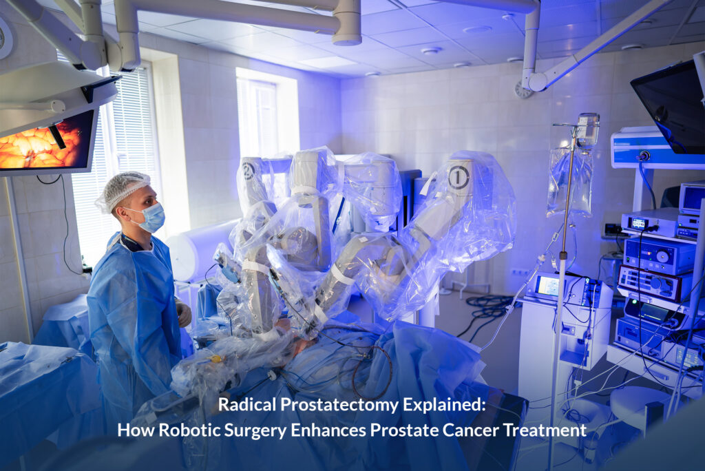
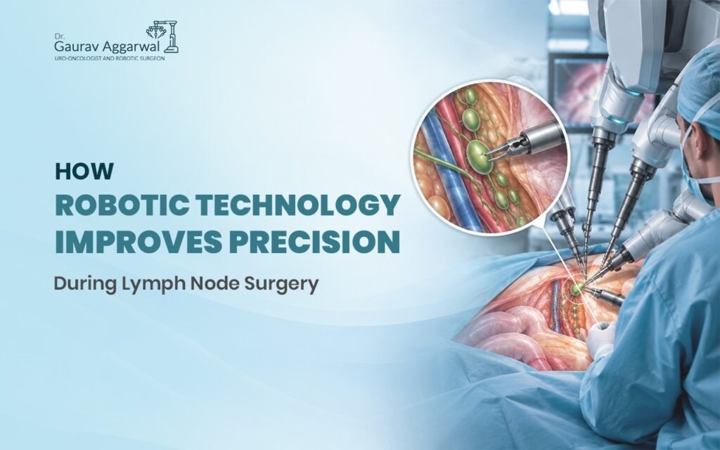
How Robotic Technology Improves Precision During Lymph Node Surgery Authored by: Dr Gaurav Aggarwal (Uro-oncologist and Robotic Surgeon)  When cancer spreads, it doesn’t travel randomly. It follows a predictable path through the lymphatic system. Surgeons have known this for decades. What changed in the last ten years is our ability to see and remove only the affected nodes—without collateral damage. Robotic technology during lymph node surgery has redrawn what’s possible. Instead of making large incisions to find and remove lymph nodes, surgeons now work through 4-5 tiny openings. The robot’s camera shows a magnified, three-dimensional view. The instruments move with precision that human hands alone cannot replicate. The result: fewer complications, faster recovery, and better cancer outcomes. This shift has been transformative for urological cancers. Prostate cancer, kidney cancer, and bladder cancer all rely on accurate lymph node assessment. Understanding whether cancer has spread to the nodes changes everything—from staging to prognosis to treatment decisions. Why Lymph Nodes Matter in Cancer Surgery The lymphatic system is the body’s early warning system. When a tumour grows, cancer cells often reach nearby lymph nodes first. If nodes are clear, the cancer is localised. If nodes are involved, the cancer has begun to spread. For urological cancers, this matters surgically: A surgeon needs to know which nodes are involved before operating Removing too many nodes causes long-term swelling (lymphoedema) Leaving involved nodes in place misses the cancer The nodes are deep—buried in fat and tissue, near vital blood vessels and organs Traditional open surgery required large incisions to access these areas safely. A surgeon could see, but only from one angle. Palpating tissue (feeling with gloved hands) helped identify nodes, but it wasn’t precise. Some nodes stayed behind. Others got removed unnecessarily. How Robotic Lymph Node Surgery Works Robotic technology doesn’t replace the surgeon. It extends the surgeon’s ability. The setup: Anaesthesia is induced, and the patient is positioned for access to the surgical area 4-5 small incisions (about 8-12 mm each) are made A camera (called an endoscope) goes through one port; instruments through the others The surgeon sits at a console 10 feet away, controlling the robot’s arms The precision advantage: The camera magnifies the area 10-12 times The image is three-dimensional, giving true depth perception The surgeon’s hand movements are scaled down—a 1cm hand movement becomes a 1-2mm instrument movement Tremor is filtered out automatically Articulated instruments bend and rotate in ways human wrists cannot What the surgeon sees: Individual lymph nodes sitting in their tissue beds Nearby blood vessels and nerves in high definition The exact plane between healthy tissue and cancerous tissue The ability to remove nodes without damaging structures nearby The Clinical Advantage Studies from urology centres across India and globally show consistent benefits: Better cancer staging: More nodes are identified and removed when indicated Staging accuracy improves, meaning treatment is based on complete information Understaging (missing nodes) drops significantly Reduced side effects: Lymphoedema rates fall because only necessary nodes are removed Infection risk is lower—smaller incisions mean less tissue trauma Nerve injury is less common because visualisation is superior Faster recovery: Patients go home 1-2 days after surgery instead of 3-5 days Return to normal activity happens in 2-3 weeks instead of 6-8 weeks Pain is minimal—small incisions mean minimal wound pain Better long-term outcomes: Cancer recurrence rates reflect improved staging and more complete node removal Quality of life improves because complications are fewer Lymph Node Surgery in Different Urological Cancers Prostate cancer: Robotic-assisted lymph node removal happens during prostatectomy Nodes in the pelvis are accessed safely through the same small incisions Precision matters because nearby nerves control sexual and urinary function Kidney cancer: Nodes around the kidney and in the retroperitoneum (behind the abdomen) are difficult to reach Robotic access allows removal without opening the belly fully The surgeon can work in a confined space without hand strain Bladder cancer: Pelvic lymph node removal is standard before removing the bladder The robotic approach is gentler on surrounding tissue Recovery is faster, which matters because bladder cancer patients often need follow-up chemotherapy Testicular cancer: Nodes in the retroperitoneum are deep and close to major blood vessels Robotic magnification reduces the risk of vascular injury Even small errors matter in this delicate region What to Expect: Before, During, and After Robotic Lymph Node Surgery Before surgery: Imaging (CT or MRI) identifies the location of nodes needing removal Blood work and cardiac clearance are standard You’ll meet the surgical team and anaesthesiologist Fasting instructions are given (usually 6 hours before surgery) During the procedure: Total time in the operating room is typically 2-4 hours depending on the extent of lymph node surgery You’re under general anaesthesia the entire time The robotic system is positioned, and small incisions are made Node removal takes place; tissue is sent to pathology Incisions are closed with dissolving stitches or clips After surgery: Recovery room monitoring for 1-2 hours Most patients go home the same day or the next morning Pain is managed with oral medication Activity restrictions are minimal—walking and light movement start immediately Shower restrictions apply to incisions until they’re sealed (usually 3-4 days) Follow-up is at 1-2 weeks, then 4-6 weeks Robotic Lymph Node Surgery: Key Advantages Magnified three-dimensional view eliminates guesswork Smaller incisions reduce pain, infection risk, and recovery time Precise node dissection means better staging and fewer side effects Nerve and blood vessel injury risk drops significantly Patients return to normal activity faster Cancer outcomes improve because staging is accurate Working With an Experienced Robotic Surgeon Not all surgeons perform robotic lymph node surgery. The learning curve is steep. A surgeon needs hundreds of cases—not dozens—before they handle complex node dissections confidently. Look for: Board certification in urology and specific robotic credentialing Documented experience with 1,000+ robotic procedures Specialisation in cancer surgery (uro-oncology) Access to current-generation robotic systems A team that follows your case before and after surgery Dr Gaurav Aggarwal, based in Newtown, Kolkata, specialises in uro-oncology with robotic precision. His experience spans over 1,500 robotic

How Robotic Technology Improves Precision During Lymph Node Surgery Read More »
What Is the Retroperitoneum? Anatomy, Organs and Functions Explained

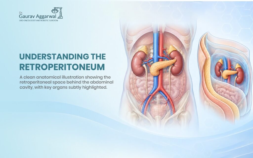
What Is the Retroperitoneum? Anatomy, Organs and Functions Explained Authored by: Dr Gaurav Aggarwal (Uro-oncologist and Robotic Surgeon)  When patients come to see me with kidney problems, prostate issues, or urological conditions, I often need to explain where things are located inside their body. That’s when I start talking about the retroperitoneum. Most people have never heard of it, and honestly, that’s understandable. It’s not an organ you can point to or something you think about unless something goes wrong. But here’s the thing—understanding what is the retroperitoneum and why it matters has helped many of my patients grasp why their condition needs the treatment it does. So let me walk you through it. What Is the Retroperitoneum, Actually? The retroperitoneum is a space. Not an organ, not a tissue—a space. Specifically, it’s the area behind your abdominal cavity that sits between the peritoneum (a membrane lining your abdomen) and the back wall of your abdomen.   Think of it this way: inside your belly, you have your intestines, stomach, liver, and other organs. These organs have a protective lining around them called the peritoneum. Everything inside that lining is called the peritoneal space. But behind that lining—behind the peritoneum—there’s another space. That’s the retroperitoneum. When I perform robotic surgery on the kidneys or prostate, I’m often working in this retroperitoneal space. This is where the important structures hide, which is why understanding the anatomy matters. What Organs Are in the Retroperitoneum? This is where it gets clinically interesting. Several vital organs live in this space, and they’re crucial to your body’s function.   The kidneys sit in the retroperitoneum on either side of your spine, one on the left and one on the right. These filter your blood and produce urine. As a urologist, the kidneys are central to what I treat.   The ureters run from your kidneys down to your bladder, carrying urine. They travel through the retroperitoneum the entire way.   The adrenal glands sit on top of each kidney. They produce hormones that regulate stress response, blood pressure, and metabolism. Major blood vessels pass through here too: The abdominal aorta (the largest artery in your body, carrying blood from your heart) The inferior vena cava (the large vein returning blood to your heart) Smaller arteries and veins branching to the organs   The pancreas sits in the retroperitoneum behind your stomach. It produces insulin and digestive enzymes. Parts of the duodenum and colon also sit retroperitoneally. These are parts of your digestive system. The psoas muscles run through the retroperitoneum along your spine, helping you flex your hip. Why Does This Matter? You might be thinking, “Okay, so these organs are back there. Why should I care where they are?” It matters because location determines treatment options. When I’m dealing with a kidney tumor or a prostate condition, the retroperitoneum means how I can access it, what structures I need to be careful around, and whether robotic surgery is the right approach.   In my practice, understanding the retroperitoneal anatomy is why we can perform minimally invasive procedures. When I use robotic surgery to remove a kidney, I’m working through the retroperitoneal space with precision. I can see the structures clearly, avoid damaging surrounding tissues, and preserve function better than with open surgery.   If you have kidney stones stuck in your ureter, they’re stuck in a retroperitoneal structure. If you have an infection in the retroperitoneum, it’s potentially serious because that space isn’t as easy to access and treat as other areas. How Does the Retroperitoneum Stay Protected? One reason retroperitoneal infections are dangerous is because this space isn’t as open as other parts of your abdomen. The organs and tissues here are enclosed by fascia—connective tissue that acts like a protective wrapper.   This fascia is called Gerota’s fascia around the kidney. It holds the kidney in place and helps contain any infection or bleeding. But it also means that if something goes wrong, it can be harder to detect and treat early because symptoms might not show up as quickly. Surgeons need to respect this anatomy. When I’m working in the retroperitoneum, I’m navigating around: Major blood vessels (you don’t want to accidentally cut the aorta) Nerve structures Existing inflammation or scarring The fascia layers This is why a surgeon’s understanding of retroperitoneal anatomy isn’t just academic—it’s essential for patient safety. Common Conditions Affecting the Retroperitoneum Over my years in practice, I’ve treated patients with various retroperitoneal conditions: Kidney stones get stuck in the ureter, which runs through the retroperitoneum. They cause intense pain and need to be removed or dissolved.   Renal cell carcinoma (kidney cancer) starts in the kidneys and may spread within the retroperitoneal space. Surgery to remove the kidney requires understanding exactly how far the cancer has spread. Retroperitoneal fibrosis is a rare condition where fibrous tissue builds up in the retroperitoneum, potentially trapping the ureters and blocking urine flow. This is serious and requires intervention. Aortic aneurysms affect the abdominal aorta, which runs through the retroperitoneum. This is life-threatening and requires urgent surgery.   Kidney infections can spread into the retroperitoneal space. Because it’s a confined space, this can become critical quickly.   Prostate conditions directly affect structures adjacent to the retroperitoneum, which is why understanding this anatomy helps with treatment planning. Retroperitoneal Surgery and Robotic Technology This is where my speciality gets interesting. When we need to surgically treat retroperitoneal conditions, we have choices. Traditional open surgery means making a large incision, opening the abdomen, and working with our hands. For some cases, this is still necessary. But robotic-assisted surgery has changed how I approach many retroperitoneal procedures. With the da Vinci robot, I have better visualization of the retroperitoneal space. The camera gives me high-definition, three-dimensional imaging. My instruments have more precision than my hands alone can achieve. This means smaller incisions, less tissue damage, faster recovery, and better outcomes for patients.   When I’m doing a robotic nephrectomy (kidney removal) for cancer, I

What Is the Retroperitoneum? Anatomy, Organs and Functions Explained Read More »

What Is a Cystectomy, and When Is It Needed?

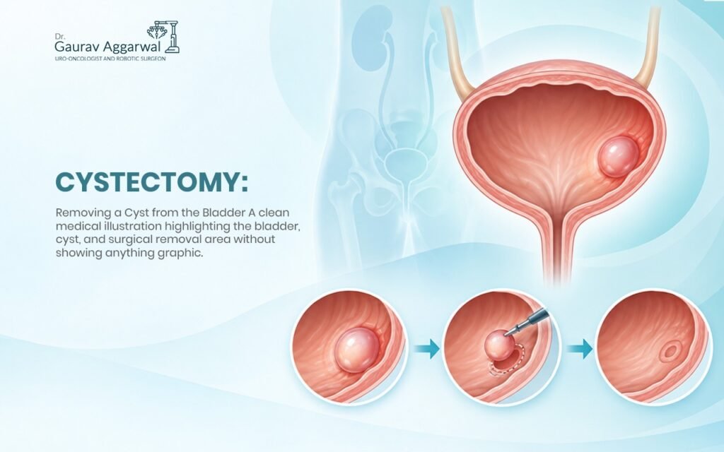
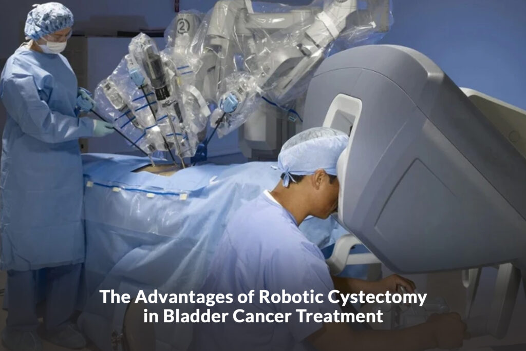
What Is a Cystectomy, and When Is It Needed? Authored by: Dr Gaurav Aggarwal (Uro-oncologist and Robotic Surgeon)  A cystectomy is the surgical removal of the bladder. If you or someone you know has been told this surgery might be necessary, you probably have questions. What does it mean to lose your bladder? How does the body handle urination afterward? Is this really the best option? This guide walks through what a cystectomy actually involves, why doctors recommend it, and what to expect. For most people, the bladder is invisible—you simply use the bathroom when you need to. But when bladder cancer develops or certain conditions damage the bladder beyond repair, removal becomes the best path forward. What a cystectomy isn’t is a small procedure. It’s a major surgery that changes how your body functions. Understanding it means understanding both the surgery itself and the real reasons your doctor is recommending it. What Is a Cystectomy: The Basic Definition What is a cystectomy in the simplest terms? It’s the surgical removal of the entire bladder. In men, this usually means removing the bladder, prostate, and seminal vesicles. In women, it means removing the bladder, uterus, ovaries, and part of the vagina. The surgeon doesn’t just take out the organ and leave nothing. They reconstruct a way for urine to leave the body. After a cystectomy, urine has nowhere to go in the traditional sense. The body needs a new path for waste. Surgeons create this through: A urostomy (ileal conduit) – using a small piece of intestine to create a new passage for urine to an opening in the abdomen A continent pouch – reshaping the intestine to create an internal reservoir that you empty through a catheter A neobladder – reconstructing a bladder-like pouch from the intestine so you can urinate more naturally, though typically you’ll need to catheterise The type of reconstruction depends on your age, health, cancer stage, and preference. Why Doctors Recommend Cystectomy The most common reason for cystectomy is bladder cancer, particularly muscle-invasive bladder cancer that has penetrated deep into the bladder wall. At this stage, removing the entire bladder gives the best chance of eliminating cancer completely. Radiation and chemotherapy might shrink tumours, but they don’t guarantee the cancer won’t return. A cystectomy removes the source. Cystectomy is also recommended for: Bladder cancer that has spread to the prostate (in men) or upper vagina (in women) Recurrent bladder cancer that keeps returning after other treatments Severe bladder damage from radiation therapy Neurogenic bladder that’s become untreatable and is causing serious infections Bladder rupture or trauma that can’t be repaired Chronic inflammation that has damaged the bladder beyond function For cancer specifically, the decision comes down to this: bladder cancer is treatable, but only if caught and treated aggressively. What a cystectomy represents is that aggressive treatment—removing the problem before it spreads further. Understanding the Surgery Itself This is major surgery requiring general anaesthesia and typically takes 3 to 5 hours. The surgeon makes an incision in the lower abdomen to access the bladder. They remove the bladder along with surrounding lymph nodes (to check for cancer spread). Then they create the new urinary system. Robotic surgery has changed how this procedure is performed. Instead of one large incision, the surgeon makes several small ones and works through a robotic system, which allows for greater precision. Robotic cystectomy reduces blood loss, shortens hospital stays, and speeds recovery compared to open surgery. During surgery, several things happen: The bladder is removed completely Nearby lymph nodes are taken and tested The urethra is carefully handled to preserve as much function as possible The new urinary pathway is created using intestinal tissue All connections are tested to ensure they work correctly Drains are placed to prevent fluid buildup The surgery is complex. Recovery takes weeks, not days. What Recovery Actually Looks Like After cystectomy, you’ll spend 2 to 4 days in the hospital. Pain management is essential for the first few weeks. Most people need 4 to 6 weeks before returning to normal activities, though full recovery takes closer to 8 to 12 weeks. During recovery: You’ll have a catheter (tube) draining urine into a collection bag – this prevents pressure on the new system while it heals Drains remove fluid from the surgical site to prevent buildup Pain medication helps manage discomfort as you heal Diet progresses gradually from liquid to solid food You’ll need to learn how to manage your new urinary system Follow-up appointments monitor healing and teach you self-care The first few weeks feel heavy. You have tubes, pain, and limited mobility. But each week improves. By 4 to 6 weeks, most people feel substantially better. By 3 months, they’re back to their regular routines—just with a different way of managing urination. Adjusting to Life After Cystectomy The permanent change is learning to manage urine without a bladder. If you have an ileal conduit (the most common reconstruction), you’ll wear a pouch on your abdomen that collects urine throughout the day. You empty it when needed—typically 4 to 6 times daily. It becomes routine, like any other daily task. With a continent pouch, you catheterise yourself through an opening in the abdomen 4 to 5 times daily. With a neobladder, you urinate more normally but may need catheterisation occasionally. The adjustment period is real. People report: Learning to manage the pouch or catheterisation effectively Adjusting to the physical sensation of something new on their body Regaining confidence in their routine and social activities Returning to work, exercise, and hobbies Adapting intimate relationships as they figure out what feels comfortable Managing occasional leakage and how to handle it discreetly Most people adapt within 2 to 3 months. The key is patience with yourself during that learning period. Why This Surgery Saves Lives For muscle-invasive bladder cancer, cystectomy offers the best survival outcomes. Patients who undergo cystectomy have higher rates of cancer-free survival compared to those who try to preserve the bladder with chemotherapy and

What Is a Cystectomy, and When Is It Needed? Read More »